The Histology and Histomorphometry Core

Bridging structure and function through precision histology 

The Histology and Histomorphometry Core provides inhouse expertise, techniques, equipment and technical innovation to facilitate Indiana Center for Musculoskeletal Health investigators from vastly different subspecialties to use histological approaches in order to assess tissue/cell level mechanisms in their developmental/physiological/disease models. The core facilitates histological and histomorphometric analyses of bone, muscle, cartilage, ligament, tendon and intervertebral disk. The core also provides consultation on study design and post-study data interpretation and provide training programs and education in all techniques covered by the core.
Services

The Histology and Histomorphometry Core offers services in the following areas:

  • Histological processing and quantitative analysis of musculoskeletal tissues:

    • Histological preparations and outcome-specific staining techniques for bone, muscle, cartilage, ligament, tendon and intervertebral disk

    • Undecalcified/decalcified hard tissues, frozen section, immunohistochemistry and special staining

    • Analytical support for histomorphometry and immunohistochemistry assessments

    • Integration with the Systems Biology & Muti-Omics Core (SBMOC) to support spatial transcriptomics of musculoskeletal tissues

  • Consistency and comparability of measurement modalities across studies

    • Quality control standards for technical procedures

    • Coordinated guidance on selection and quality management of appropriate preclinical and clinical assessments

    • Centralized assessment of outcomes

  • Consultation on study design and post-study data interpretation

  • Training and education in histological analysis

    • Annual training sessions for faculty, fellows and graduate students

    • Two one-day trainings provided each year focused on one of the approaches utilized by the core

    • One-on-one training with laboratory supervisors or core director on request

  • Technical personnel professional development training

View pricing, policies and protocols

Schedule in iLab

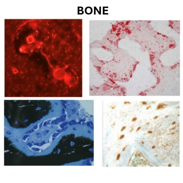
bone histology examples
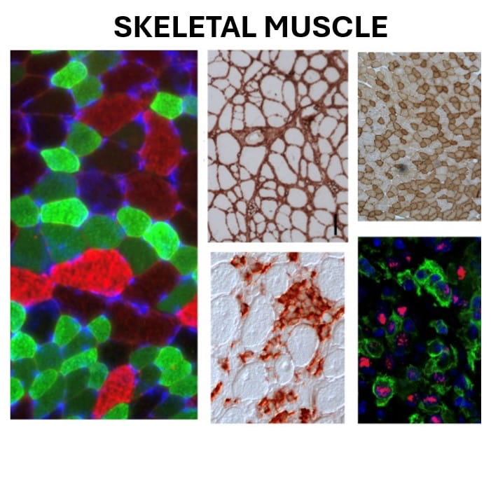
skeletal muscle examples
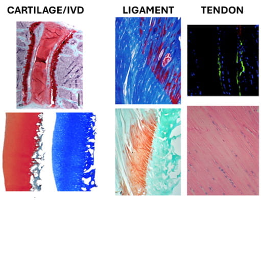
histology of cartilage, ligament and tendon

Leadership

19291-Plotkin, Lilian

Lilian I. Plotkin, PhD

Professor of Anatomy, Cell Biology & Physiology

Core Director

Read Bio

5035-Allen, Matthew

Matthew R. Allen, PhD

Professor of Anatomy, Cell Biology & Physiology

Core Co-Director

Read Bio

Acknowledgment Policy

In all cases, the core requests that the following statement be placed in an acknowledgements section of any published work that utilizes core facilities: 

“This work was supported by the Histology Core of the Indiana Center for Musculoskeletal Health at IU School of Medicine and the Indiana Clinical Translational Sciences Institute (CTSI).” 


Publications

Srivastava, S., Jiang, J., Misra, J. et al. Asparagine bioavailability regulates the translation of MYC oncogene. Oncogene 41, 4855–4865 (2022). https://doi.org/10.1038/s41388-022-02474-9 

Essex AL, Huot JR, Deosthale P, Wagner A, Figueras J, Davis A, Damrath J, Pin F, Wallace J, Bonetto A, Plotkin LI. Triggering Receptor Expressed on Myeloid Cells 2 (TREM2) R47H Variant Causes Distinct Age- and Sex-Dependent Musculoskeletal Alterations in Mice. J Bone Miner Res. 2022 Jul;37(7):1366-1381. doi: 10.1002/jbmr.4572. Epub 2022 Jun 7. PMID: 35575023; PMCID: PMC9307075. 

Moraes de Lima Perini M, Valuch CR, Dadwal UC, Awosanya OD, Mostardo SL, Blosser RJ, Knox AM, McGuire AC, Battina HL, Nazzal M, Kacena MA, Li J. Characterization and assessment of lung and bone marrow derived endothelial cells and their bone regenerative potential. Front Endocrinol (Lausanne). 2022 Aug 31;13:935391. doi: 10.3389/fendo.2022.935391. PMID: 36120459; PMCID: PMC9470942. 

Dudakovic, A., Jerez, S., Deosthale, P.J. et al. MicroRNA-101a enhances trabecular bone accrual in male mice. Sci Rep 12, 13361 (2022). https://doi.org/10.1038/s41598-022-17579-0  

Sakharkar M, Rappazzo CG, Wieland-Alter WF, Hsieh CL, Wrapp D, Esterman ES, Kaku CI, Wec AZ, Geoghegan JC, McLellan JS, Connor RI, Wright PF, Walker LM. Prolonged evolution of the human B cell response to SARS-CoV-2 infection. Sci Immunol. 2021 Feb 23;6(56):eabg6916. doi: 10.1126/sciimmunol.abg6916. PMID: 33622975; PMCID: PMC8128290. 

Wang J, Pajulas A, Fu Y, Adom D, Zhang W, Nelson AS, Spandau DF, Kaplan MH. γδ T Cell‒Mediated Wound Healing Is Diminished by Allergic Skin Inflammation. J Invest Dermatol. 2022 Oct;142(10):2805-2816.e4. doi: 10.1016/j.jid.2022.03.012. Epub 2022 Apr 1. PMID: 35378112; PMCID: PMC9509419. 

Hsiao YT, Manikowski KJ, Snyder S, Griffin N, Orr AL, Hulsey EQ, Born-Evers G, Zukosky T, Squire ME, Hum JM, Metzger CE, Allen MR, Lowery JW. NMUR1 in the NMU-Mediated Regulation of Bone Remodeling. Life (Basel). 2021 Sep 29;11(10):1028. doi: 10.3390/life11101028. PMID: 34685399; PMCID: PMC8538501. 

Eaton MS, Weinstein N, Newby JB, Plattes MM, Foster HE, Arthur JW, Ward TD, Shively SR, Shor R, Nathan J, Davis HM, Plotkin LI, Wauson EM, Dewar BJ, Broege A, Lowery JW. Loss of the nutrient sensor TAS1R3 leads to reduced bone resorption. J Physiol Biochem. 2018 Feb;74(1):3-8. doi: 10.1007/s13105-017-0596-7. Epub 2017 Oct 10. PMID: 29019082. 

Moraes de Lima Perini M, Valuch CR, Dadwal UC, Awosanya OD, Mostardo SL, Blosser RJ, Knox AM, McGuire AC, Battina HL, Nazzal M, Kacena MA, Li J 2022 Characterization and assessment of lung and bone marrow derived endothelial cells and their bone regenerative potential. Front Endocrinol 13:935391. doi: 10.3389/fendo.2022.935391 

Jacobs C, Shah S, Lu WC, Ray H, Wang J, Hockaden N, Sandusky G, Nephew KP, Lu X, Cao S, Carpenter RL. HSF1 Inhibits Antitumor Immune Activity in Breast Cancer by Suppressing CCL5 to Block CD8+ T-cell Recruitment. Cancer Res. 2024 Jan 16;84(2):276-290. doi: 10.1158/0008-5472.CAN-23-0902. PMID: 37890164; PMCID: PMC10790131. 

Roy Gitanjali, Syed Rameesha, Lazaro Olivia, Robertson Sylvia, McCabe Sean D., Rodriguez Daniela, Mawla Alex M., Johnson Travis S., Kalwat Michael A. (2024) Identification of type 2 diabetes- and obesity-associated human β-cells using deep transfer learning eLife 13:RP96713. https://doi.org/10.7554/eLife.96713.1 

Ghobashi AH, Lanzloth R, Ladaika CA, Masood A, O'Hagan HM. Single-Cell Profiling Reveals the Impact of Genetic Alterations on the Differentiation of Inflammation-Induced Murine Colon Tumors. Cancers (Basel). 2024 May 28;16(11):2040. doi: 10.3390/cancers16112040. PMID: 38893159; PMCID: PMC11171101. 

Ladaika CA, Ghobashi AH, Boulton WC, Miller SA, O'Hagan HM. LSD1 and CoREST2 Potentiate STAT3 Activity to Promote Enteroendocrine Cell Differentiation in Mucinous Colorectal Cancer. Cancer Res. 2025 Jan 2;85(1):52-68. doi: 10.1158/0008-5472.CAN-24-0788. PMID: 39365378; PMCID: PMC11695186. 

Mohan G, Khan I, Neumann CR, Jorge MD, Ahmed S, Hulsman L, Sinha M, Gordillo GM, Sen CK, Hassanein AH. Topical tissue nanotransfection of Prox1 is effective in the prophylactic management of lymphedema. Mol Ther Nucleic Acids. 2024 Jan 18;35(1):102121. doi: 10.1016/j.omtn.2024.102121. PMID: 38333673; PMCID: PMC10850858. 

Yadav AK, MacNeill JJ, Krylov A, Ashrafi N, Mimi RA, Saxena R, Liu S, Graham SF, Wan J, Morral N. Sex- and age-associated factors drive the pathophysiology of MASLD. Hepatol Commun. 2024 Aug 26;8(9):e0523. doi: 10.1097/HC9.0000000000000523. PMID: 39185904; PMCID: PMC11357696.