Pediatric pathology
Author(s)
Kendall Wilson, MD
Erin Rudzinski, MD
Summary of clinical history
The patient is a 13-year-old female who presented with new acute right upper quadrant pain and nausea. CT imaging revealed a cystic fluid collection measuring up to 4.5 cm in the anterior abdomen, abutting the urinary bladder and ascending toward the umbilicus. Visualization intraoperatively revealed a cystic structure attached to the terminal ileum, which was completely resected.
Gross findings
The ileocecectomy specimen included an intact bulging mass measuring 5.5 x 4.5 x 4.1 cm at the stapled ileal resection margin. Further examination revealed a unilocular cystic cavity filled with white mucoid material with a wall thickness averaging 0.1 cm. The mass was grossly adherent to the terminal ileum.
Microscopic findings
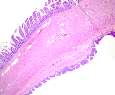
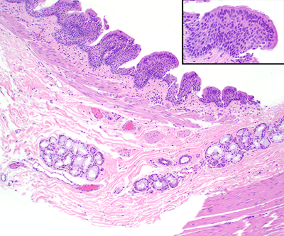
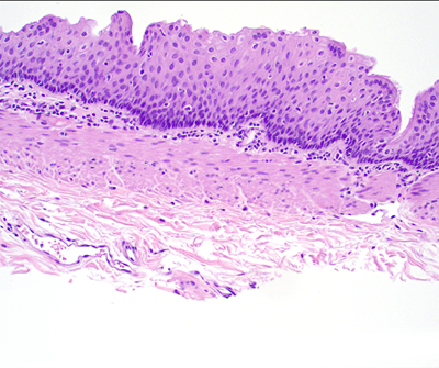
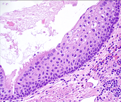
Cystic structure with a discrete muscular lining and an epithelial surface variably composed of nonkeratinizing squamous epithelium and ciliated, pseudostratified columnar epithelium with subepithelial seromucinous glands.
Click any image for larger version
Figure 1. Histology demonstrates the cyst (bottom) directly attached to the intestinal wall (4x).
Figure 2. Closer view of the cyst shows ciliated pseudostratified columnar epithelium with muscularis mucosa and submucosal mucinous glands (10x). Hyaline cartilage is notably absent. The inset at the top right provides a closer view of the cilia (40x).
Figure 3. A different area of the cyst lining shows non-keratinizing stratified squamous mucosa with a thinned, but distinct underlying muscularis propria(20x).
Figure 4. This area of the cyst lining shows a clear transition between the ciliated pseudostratified columnar epithelium and the stratified squamous mucosa (40x).
Immunohistochemical findings
N/A


