Pediatric Pathology
Authors
Harini Venkatraman Ravisankar, MBBS
Erin Rudzinski, MD
Summary of clinical history
A 10-year-old male patient noticed a small mass on his right upper arm two years ago. Over time, the mass increased in size to approximately 1.5 cm and became tender to touch. Surgical excision of the mass was performed.
Gross findings
The specimen consisted of fragmented, tan-yellow, firm, lobulated fibroadipose tissue, measuring a total of 2.4 × 2.1 × 0.6 cm. One of the fragments included an attached ellipse of skin.
Microscopic findings
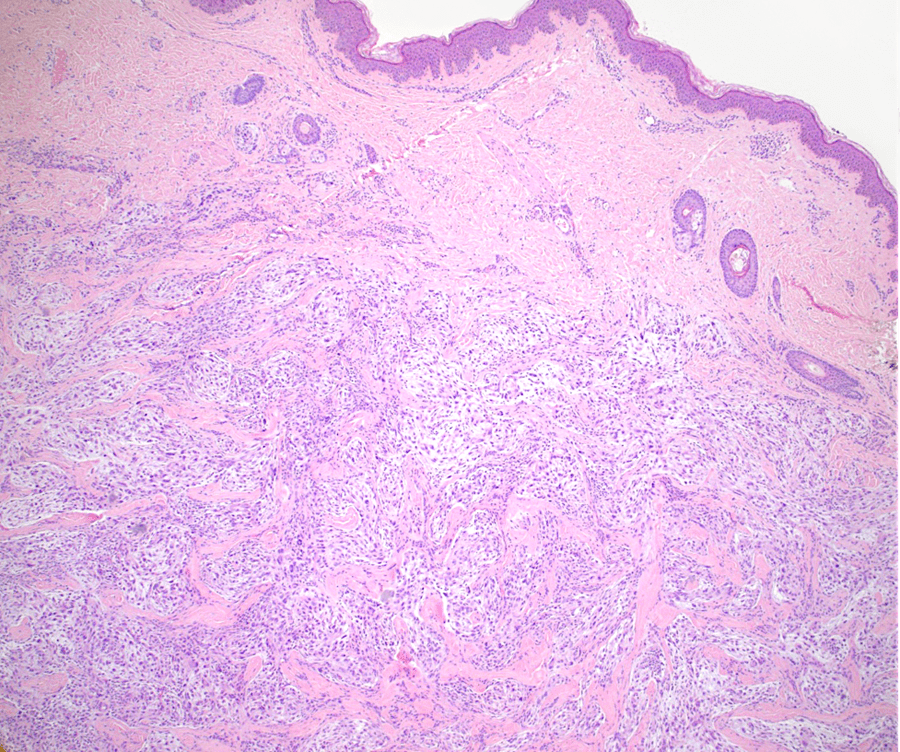
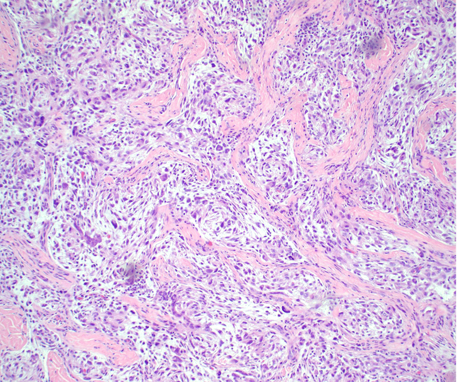
- Spindled to epithelioid cells arranged in whorls and fascicles against a myxoid background
- The cells are present in the dermis
- Dense bundles of collagen separate the cells into nodules
- Variable nuclear atypia
Click any image for larger version.
Figure 1: H&E, 40x- Dermal nodule with lobulated nests of spindle-to-epithelioid cells separated by dense collagen
Figure 2: H&E, 100x- Individual cells are spindle-to-epithelioid against a myxoid stroma
Immunohistochemical findings
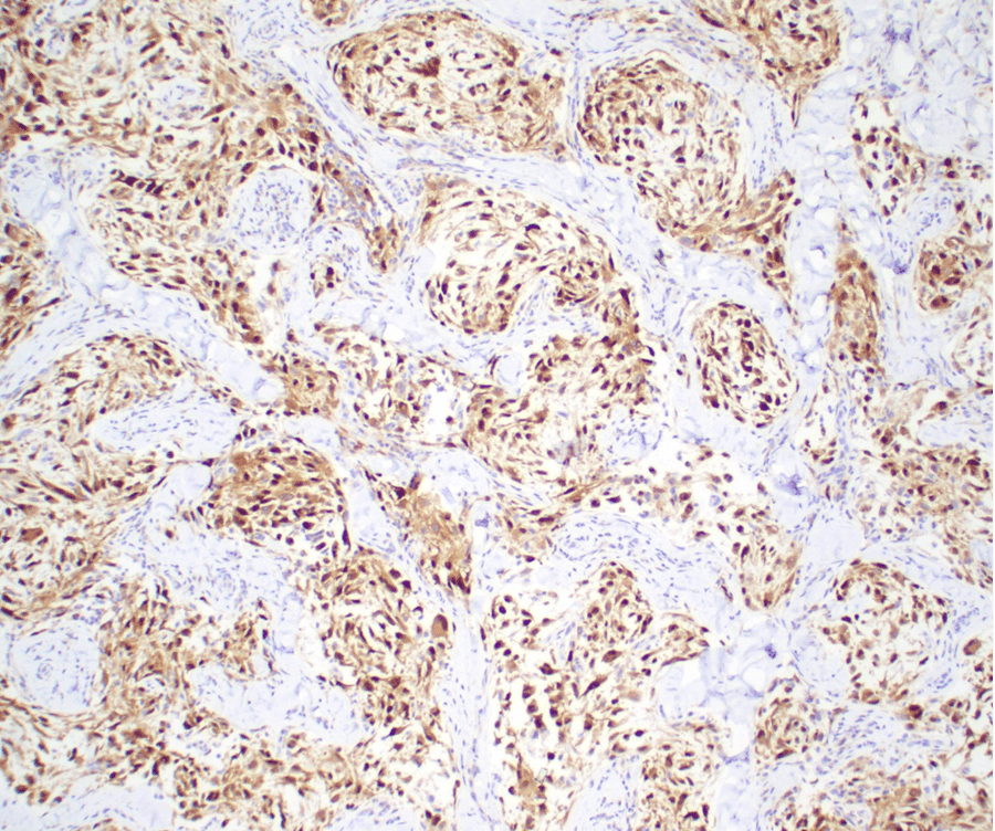
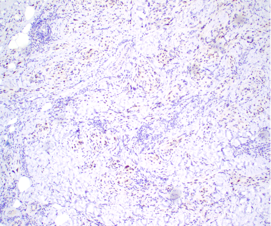
- Positive stains: PGP9.5 and MiTF (subset)
- Negative stains: S100, GFAP and Melan A
Figure 3: IHC, 100X- The cells are highlighted by PGP9.5 immunohistochemical stain
Figure 4: IHC, 100X- A subset of the cells highlighted by MiTF immunohistochemical stain


