Medical Imaging Research Institute

Cardiovascular Imaging Research Center

The Cardiovascular Imaging Research Center (CIRC) at Medical Imaging Research Institute (MIRI) advances cardiovascular research and discovery through the design, development and deployment of advanced imaging tools and translational research.

Exterior image of the Research II building on IU Indianapolis' campus.

Our objective

CIRC investigators study various forms of diseases that affect the heart muscle, with a major focus on ischemic heart disease (IHD), a class of heart disease that stems from reduced oxygen supply to the heart muscle. IHD is the most common cause of death in western countries.

Our scientists aspire to better understand heart disease from the standpoint of structural, compositional and functional changes to the heart in the early, progressive and terminal stages of the disease using an integrated multidisciplinary approach.

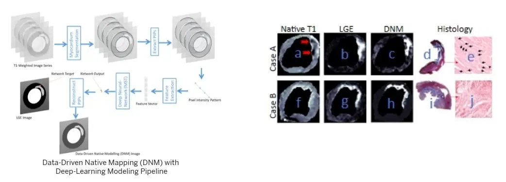
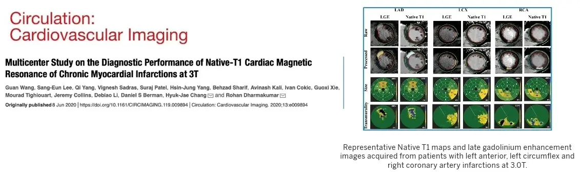
We conduct these studies using sophisticated imaging technologies, particularly MRI and PET, biomedical engineering methods, molecular immunopathological analyses, multi-omics and informatics.

Identifying biomarkers to predict progression of heart disease

Funded by multiple agencies including the National Institutes of Health and the American Heart Association, CIRC scientists are investigating novel tools for early diagnosis of disease through the identification of biomarkers for non-invasive and reliable detection of myocardial injury in the progressive phase of disease advancement, that will aid in early diagnosis of disease timely intervention strategies.

A graphic of blue circles connecting to a larger, darker blue circle. The center represents integrated therapeutics for IHD. The circles around it are physics and engineering, pathology/cell biology mechanisms, AI-driven investigations, biophysics physiology, cardiac imaging, and clinical trials.

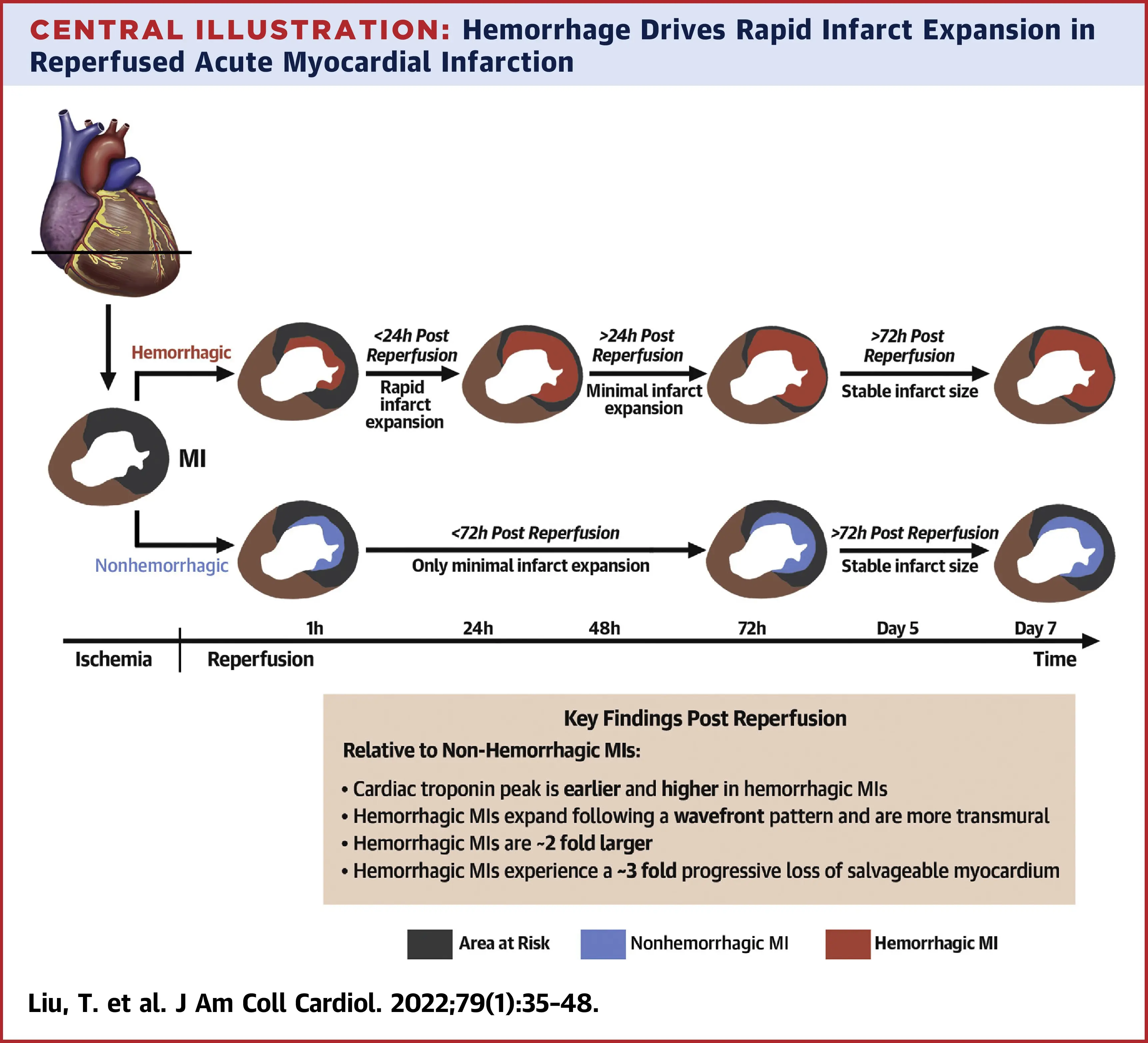
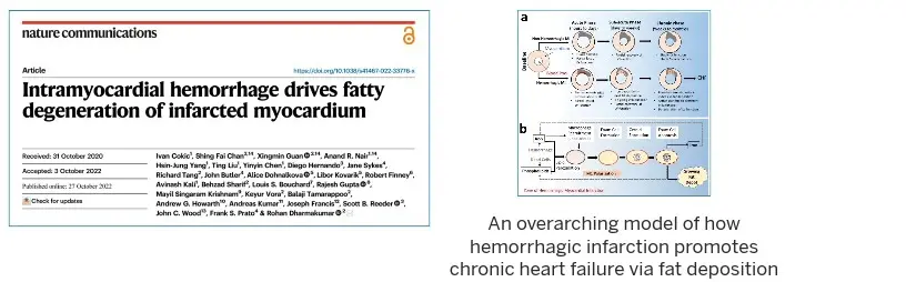
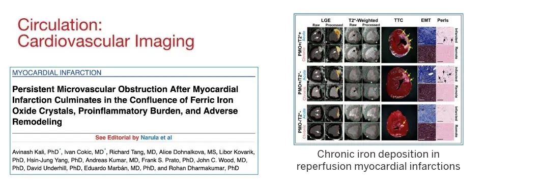
In November 2022, our landmark study published in Nature Communications showed that residual iron following heart attacks plays a causative role in chronic heart failure, through the formation of fatty tissues in the heart.

That study formed a central basis in the development of the first ever clinical classification of heart attacks that can help inform clinicians on the best treatment options based on the stage of heart damage. This classification was officially adopted by the Canadian Cardiovascular Society in 2024 and has been well received by the international cardiovascular research community. It also led to the first-in-human FDA approved trial to treat hemorrhagic myocardial infarction.

In September 2025, another major milestone was achieved by our scientists enabling the detection of hemorrhagic myocardial infarction based on a blood test. This test is expected to open new opportunities for novel therapies for patients developing hemorrhagic myocardial infarction (the worst form of heart attacks) and to empower imaging approaches for deep tissue characterization.

Areas of focus

The artificial intelligence (AI)-driven investigations into IHD are tightly interwoven with cardiac imaging as well as the use of AI to develop predictive models that can be embedded in detection systems. Key advances and ongoing work include:

  • Amplification of native T1 image contrast via data-driven native contrast mapping for rapid visual detection of chronic myocardial infarction without contrast media
  • Automated detection of and characterization of coronary artery collaterals with deep learning
  • Predictive modeling outcomes in MI patients using explainable-AI

Featured Publication

Enabling Reliable Visual Detection of Chronic Myocardial Infarction with Native T1 Cardiac MRI Using Data-Driven Native Contrast Mapping. Yousef K, Zhang X, Yoosefian G, Chen Y, Fai-Chan S, Yang HJ, Vora K, Howarth AG, Kumar A, Sharif B, Dharmakumar R. Radiol Cardiothorac Imaging. 2024 July 18:e230338

graphic from this publication shows data driven native mapping with a deep-learning modeling pipeline

Discovery work is an important aspect of our investigations into ischemic heart disease. In addition to developing pathophysiological insights based on noninvasive and invasive imaging standards, the program aspires to develop novel biophysical and physiological insights into cardiac function. Active investigations along with major contributions include:

  • Time dependence of myocardial oxygenation throughout cardiac cycle in health and IHD
  • Effect of arterial blood gases on red blood cells, vascular endothelium and cardiomyocytes
  • Modulation of mitochondrial function in response to temperature changes
  • Mechanisms modulating rest perfusion in the heart

Our contributions to date include:

  • Delineation of cyclic blood volume and oxygenation changes at rest and during pharmacological stress with cardiac MRI
  • Hypercapnia as a potent coronary vasodilator with PET and MRI
  • Rest perfusion in myocardial infarction with cardiac MRI

Featured Publications

Intramyocardial Hemorrhage and the “Wave-front” of Reperfusion Injury. Liu T, Howarth AG, Nair AR, Chen Y, Yang HJ, Ren D, Tang R, Sykes J, Kovacs MS, Dey D, Slomka P, Wood JC, Finney R, Zeng M, Prato FS, Francis J, Berman DS, Shah PK, Kumar A, Dharmakumar R.

Influence of Myocardial Hemorrhage on Staging of Reperfused Myocardial Infarctions With T2 Cardiac Magnetic Resonance Imaging: Insights Into the Dependence on Infarction Type With Ex Vivo Validation
. Wang G, Yang HJ, Cokic I, Qi Y, Xie G, Francis J, Li S, Dharmakumar R. J Am Coll Cardiol Img. 2019 Apr, 12 (4) 693–703.

Influence of Myocardial Hemorrhage on Staging of Reperfused Myocardial Infarctions With T2 Cardiac Magnetic Resonance Imaging: Insights Into the Dependence on Infarction Type With Ex Vivo Validation

We are leading multiple clinical trials across the globe with multi-center investigations into myocardial infarction, novel IHD diagnosis methods, and AI applications in IHD. Our primary clinical trial focus areas include:

  • Interventional trials fueled by novel therapeutic target
  • Diagnostics trials for detection of disease
  • Predictive models to identify most vulnerable IHD patients

Our contributions to date include:

  • Identification of biomarkers that enable detection of hemorrhagic myocardial infarctions
  • Modulation in myocardial oxygenation in response to hypercapnia, a buildup of carbon dioxide in the blood stream
  • Detection of chronic myocardial infarction without exogenous contrast media

Featured Publication

In-Hospital Mortality in Hemorrhagic Myocardial Infarction. Vora KP, Kalra A, Shah CD, Bhatt K, Kumar A, Pandya T, Poptani V, Chan SF, Singh D, Jambunathan N, Subramanian R, Chen Z, Kanakasabai S, Finney R, Desai A, Kreutz R, Kovacs RJ, Raman SV, Bhatt DL, Dharmakumar R. NEJM Evidence. 2025 Sep;4(9)

graphic from this publication shows representative native T1 maps and late gadolinium enhancement images from patients with left anterior, left circumflex nad right coronary artery infarctions at 3.0T

CIRC’s integrated therapeutics research program builds on the knowledge gained from the multifaceted interdisciplinary investigations into ischemic heart disease, to develop:

  • Targeted therapeutics that will maximize cardio-protection in myocardial infarction through both in vitro and in vivo experiments. 
  • Non-invasive diagnostic tools that complement imaging diagnostics and allow for development of timely implementation of mitigation strategies.
  • Point-of-care devices that improve the efficacy of disease management.

Developing personalized targeted treatment and integrating the tools listed above have the potential to radically change the standard of care in IHD.

Featured Publication

Intramyocardial Hemorrhage Drives Fatty Degeneration of Infarcted Myocardium. Cokic I, Chang SF, Nair AR, Guan X, YangHJ, Liu T, Chen Y, HernandoD, Sykes J, Tang R, Butler J, Dohnalkova A, Kovarik L, Finney R, Kali A, Sharif B, Bouchard L, Gupta R, Krishnam MS, Vora K, Tamarappoo B, Howarth A, Kumar A, Francis J, Reeder SB, Wood JC, Prato FS, Dharmakumar R. Nat Commun13, 6394 (2022).

graphic from this publication shows an overarching model of how hemorrhagic infarction promotes chronic heart failure via fat deposition

We have a robust team of faculty investigators who specialize in pathology, cell/molecular biology with an emphasis on multi-omics studies. We are deliberate in our investigations into understanding the pathological changes the heart undergoes during the progression of ischemic heart disease. We conduct research into fundamental processes driving pathological changes observable across multitudes of spatial scales [visual (cm), noninvasive imaging (mm), histology (um), electron microscopy (nm)]. We use observations and fundamental cell biology paradigms to develop and refine mechanisms that drive pathophysiological processes. Our team is focused on various aspects of heart disease which broadly span:

  • Biomarker development
  • Drug discovery and testing
  • Cardiac immunopathology
  • Heart-brain interaction

Salient areas of research include: 

  • Identifying a set of time-dependent signaling cascade events initiated by the red-blood-cell derived iron to drive fat depositions in the myocardial infarction territory during intracardial hemorrhage using lipidomic and proteomic analysis
  • Neuromuscular interactions following reperfused myocardial infarction studying the immune responses in myocardial infarction
  • Development of bioinformatic tools from immunopathological sections to evaluate disease progression as well as efficacy of therapeutics

Our contributions to date include the:

  • Inception of the “Crystal hypothesis,” driving post infarction heart failure
  • Processes driving lipomatous metaplasia of infarcted myocardium, an adverse physiological response to myocardial infarction
  • Delineation of rapid infarct expansion following reperfusion therapy in the setting of hemorrhagic myocardial infarction
  • Novel testing of iron-chelation therapy to mitigate chronic heart failure

Featured Publication

Persistent Microvascular Obstruction Culminates in the Confluence of Iron Oxide Nanocrystal Formation, Proinflammatory Burden and Adverse Left Ventricular Remodeling in Chronic Myocardial Infarction. Kali A, Cokic I, Tang R, Dohnalkova A, Kovarik L, Yang HJ, Kumar A, Prato FS, Wood JC, Underhill D, Marban E, Dharmakumar R. Circulation: Cardiovascular Imaging 2016 Nov;9(11). pii: e004996

graphic from this publication shows chronic iron deposition in reperfusion myocardial infarctions

Physics and engineering play an integral part in the conceptualization, design and development of algorithms, tools and instrumentation that support multiple areas of IHD investigations. Key areas of focus in physics for our team include:

  • Magnetic resonance relaxometry
  • Electrical conduction in tissue
  • Fluid mechanics in small and large blood vessels

Engineering efforts are focused on:

  • Advanced imaging and reconstruction strategies
  • Development and optimization of image contrast
  • Development of devices and instrumentation to monitor and deliver therapies

Select Publications (past five years)


Select Funding (past five years)

  • NIH 1R01-HL147133-01 (MPI: Dharmakumar R (contact); Francis J)
    Mechanistic Insights to a Translatable Therapy for Acute Reperfused Hemorrhagic Myocardial Infarctions

  • NIH R01-HL148788 (PI: Dharmakumar R)
    Accurate, Needle-Free, MRI-based Detection of Ischemic Heart Disease without Contrast Agents

  • NIH R01-HL133407 (PI: Dharmakumar R)
    Developing a MRI-guided Disease-Modifying Therapy for Post Infarction Chronic Heart Failure

  • NIH R01-HL136578 (PI: Dharmakumar R)
    An Accurate Non-Contrast-Enhanced Cardiac MRI Method for Imaging Chronic Myocardial Infarctions: Technical Developments to Rapid Clinical Validation


Select Patents (past five years)

  • Dharmakumar R et al. Robust Myocardial Blood Oxygen Level Dependent Magnetic Resonance Imaging with Improved Patient Comfort. (Filed: Sep 30, 2016; Granted: Sept 9, 2022) US11,445,912 B2Dharmakumar R. Methods for reducing ischemia-reperfusion injury. (Filed: Aug 05, 2014; Granted: Nov 11, 2019). US2016/0166606 A1

  • Dharmakumar R et al. Assessment of Iron Deposition Post Myocardial Infarction as a Marker of Myocardial Hemorrhage. USPTO Application Number: 14/125,307 filed on June 13, 2011. Assignee: Cedars-Sinai Medical Center. Awarded February 27, 2020.

  • Dharmakumar R et al. Assessment of Iron Deposition Post Myocardial Infarction as a Marker of Myocardial. Continuation in Part, Application Number: 15/064,817 filed on March 9, 2016. Assignee: Cedars-Sinai Medical Center. Awarded February 27, 2020.

  • Dharmakumar R, et al. Assessment of Coronary Heart Disease with Carbon Dioxide. USPTO Application Number: (Filed: May 07, 2012; Granted: Sept 13, 2022) US 11,439,309 B2


A key aspect of our imaging focus is the technical development of methodologies to overcome unmet needs in caring for ischemic heart disease patients. Current emphases include:

  • Rapid, contrast-free (native) imaging
  • Detection and optimization of image contrast
  • Fast/robust time-resolved 3D cardiac MRI in the presence of motion (cardiac, respiratory and flow) for myocardial tissue characterizations
  • Joint MRI/PET acquisitions for multi-model characterization of IHD

The center has developed MRI technologies that allow for assessment of changes in heart in response to ischemic heart disease while free breathing, where the patient just needs to lay down and they don't need to hold their breath. Our goal is to empower these imaging approaches to decipher novel pathophysiological insight into ischemic heart disease.

We have made several seminal contributions to the field, including:

  • Imaging oxygenation changes in the heart
  • Arterial CO2 as an alternate stressor to pharmacological agents for stress imaging
  • Enabled imaging of myocardial infarctions without contrast agents
  • Developed imaging methods exposing mechanisms accelerating chronic heart failure
  • Jointly characterized ischemic myocardium through simultaneous magnetic resonance imaging (MRI)- and positron emission tomography (PET) in myocardial obstructive coronary artery disease and myocardial infarction 
  • Developed novel imaging markers of heart failure and ventricular arrhythmia in IHD

Featured Publication

Accurate Needle-Free Assessment of Myocardial Oxygenation for Ischemic Heart Disease. Yang HJ, Ilkary O, Dey D, Sykes J, Klein M, Butler J, Kovacs MS, Sobczyk O, Cokic I, Slomka P, Bi X, Li D, Tighiouart M, Prato FS, Tsaftaris SA, Fisher JA, Dharmakumar R. Science Translational Medicine. 2019 May 29;11(494).

Past and present collaborators

Midwest

Rajesh Gupta, MD, University of Toledo
Jay Traverse, MD, Minneapolis Heart Institute
Grant Reed, MD, Cleveland Clinic
Scott Reeder, MD/PhD, University of Wisconsin
Diego Hernando, PhD, University of Wisconsin

Northeast

Nikolas Frangogiannis, MD, Albert Einstein University
Ankur Kalra, MD, SUNY Upstate Medical University

South

Joseph Francis, PhD, Louisiana State University

West

Daniel Berman, MD, Cedars-Sinai MC
Louis Bouchard, University of California, Los Angeles (UCLA)
Sumeet Chugh, MD, Cedars-Sinai MC
Damini Dey, PhD, Cedars-Sinai MC
Alice Dohnalkova, Pacific Northwest National Laboratory (PNNL)
Jennifer Van Eyk, PhD, Cedars-Sinai MC
Libor Kovarik, PhD, Pacific Northwest National Laboratory (PNNL)
Debiao Li, PhD, Cedars-Sinai MC
Daniel Luthringer, MD, Cedars-Sinai MC
Eduardo Marban, MD/PhD, Cedars-Sinai MC
Prediman K Shah, MD, Cedars-Sinai MC
Piotr Slomka, PhD, Cedars-Sinai MC
Balaji Tamarappoo, MD/PhD, Mayo Clinic
David Underhill, PhD, Cedars-Sinai MC
John Wood, MD/PhD, University of Southern California
Hsin-Jung Yang, Cedars-Sinai MC

Canada

Joseph A Fisher, University of Toronto
Andrew Howarth, MD/PhD, University of Calgary
Andreas Kumar, MD, Northern Ontario School of Medicine
Michael Kovacs, PhD, Lawson Health Research Institute
Frank Prato, PhD, Lawson Health Research Institute
James White, MD, University of Calgary

United Kingdom

Vanessa Ferreria, MD/PhD, Oxford University
Pier Masci, MD, Kings College
Mayooran Shanmuganathan, MD/PhD, Oxford University
Sotirios Tsaftaris, University of Edinburgh, UK

China

Yang Qi, MD/PhD, Beijing, China