Researchers at Indiana University School of Medicine Department of Urology are learning new ways to diagnose and treat prostate cancer through prostate-specific membrane antigen positron-emission tomography, or PSMA-PET, imaging. PSMA is an antigen that attaches to the cancer. Used as a small, molecular tracer during PET-imaging, it causes the cancer to “glow” during imaging. This allows physicians to get a clearer view of the cancer and better diagnose patients. Researchers are studying the use of PSMA-PET imaging in all stages of prostate cancer—from initial screenings and active surveillance to treatment of early recurring and metastatic prostate cancer.

Initial Screening and Diagnosis
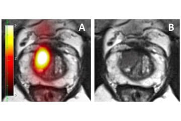
PSMA-PET imaging is used to screen for and diagnose prostate cancer. Researchers completed a pilot trial that found PSMA-PET imaging (figure A) successfully showed better images of prostate cancer than magnetic resonance imaging (MRI) alone (figure B). Another clinical trial is now underway to further study the use of PSMA-PET imaging when screening for and diagnosing prostate cancer.
Primary Treatment
Researchers are also developing ways to use the PSMA tracer to not only highlight where tumors on the prostate are located, but also kill cancerous tumors and identify where that cancer may have spread. This helps urologists treat prostate cancer using PSMA. PSMA can also be used to aid other treatments, such as high intensity focused ultrasound (HIFU) or robotic prostatectomy. In these treatments, PSMA can help urologists get a more detailed view of the cancer and have a more direct target for these treatments.
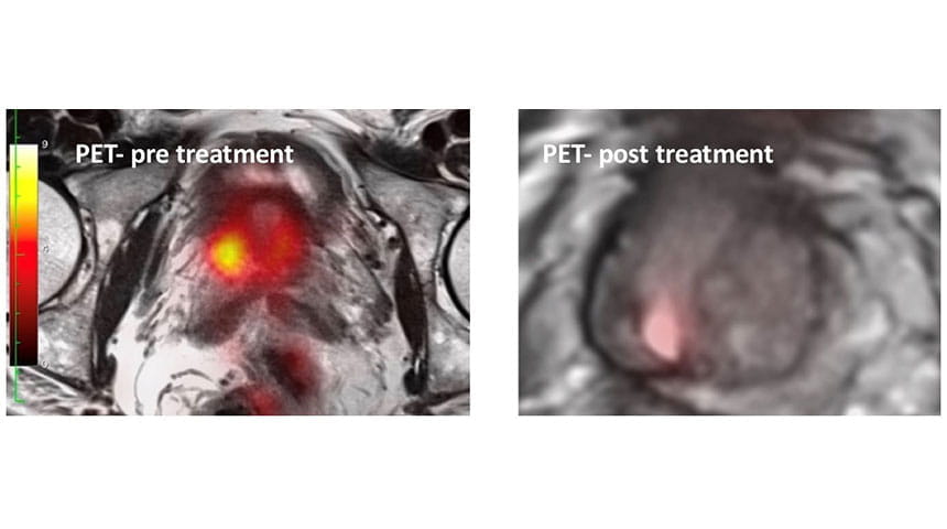
In the above figures, the prostate cancer was diagnosed with a PSMA-PET scan (left image) and then treated with high intensity focused ultrasound (HIFU). Six months after HIFU treatment, the PSMA-PET was negative (right image). The prostate-specific antigen (PSA) level went from 10.0 to 1.4.

Early Recurrence
Researchers are also studying the use of PSMA to detect recurring prostate cancer. When screening for prostate cancer, physicians monitor a patient’s PSA level. By using PSMA-PET imaging, the PSMA tracer can attach to the recurring cancer and highlight it in the imaging so they can see the cancer better than through an MRI alone.
Metastatic
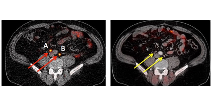
PSMA also is used to find aggressive prostate cancer that has spread outside the prostate. Because PSMA is a tracer that attaches to prostate cancer, it will also attach to cancer that may have spread to other parts of the body (see figure below). The PET imaging then shows urologists where the cancer has spread and helps them determine the best treatment options. The images below show an example of positive PSMA-PET images (left) compared with 11C-acetate-PET (right) in a prostate cancer patient with recurrent prostate cancer to lymph nodes.


