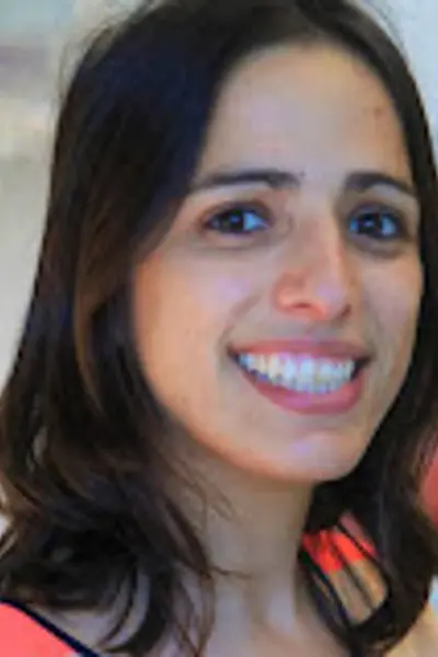
Dagan Segal, PhD
Assistant Professor of Biochemistry, Molecular Biology & Pharmacology
- dagsegal@iu.edu
- Phone
- (812) 856-1207
- Address
-
MYERS HALL 200
MSCI
BL
Bloomington, IN
Bio
Dr. Dagan Segal earned a B.S. in Cellular and Molecular Biology from the University of Texas at Austin, an M.S. in Biology from the Technion–Israel Institute of Technology, and a Ph.D. in Biology from the Weizmann Institute of Science. Her doctoral research under the mentorship of Dr. Ben-Zion Shilo examined the cellular mechanisms governing cytoskeletal dynamics in the Drosophila embryo. Dr. Segal completed postdoctoral training at the University of Texas Southwestern Medical Center in Dallas, where she worked with Drs. Gaudenz Danuser and James Amatruda to investigate Ewing sarcoma cell morphology, signaling, and plasticity in zebrafish and 3D culture models. In 2026, she joined the Indiana University School of Medicine–Bloomington as an Assistant Professor. Supported by an NCI K99/R00 Pathway to Independence Award, Dr. Segal’s research seeks to understand how tumor cells adapt to distinct tissue environments and how context-dependent changes in cell state contribute to cancer survival and progression.
Key Publications
1. Segal D, Wang X, Mazloom-Farsibaf H, Rajendran D, Butler E, Chen B, Chang B-J, Ahuja K, Perny A, Bhatt K, Reed DK, Castrillon D, Lee J, Jeffery E, Wang L, Williams NS, Rajaram S, Fiolka RP, Skapek SX, Hon GC, Amatruda J, Danuser G (2024). Caveolin-1 regulates context-dependent signaling and survival in Ewing Sarcoma. bioRxiv (preprint). doi: https://www.biorxiv.org/content/10.1101/2024.09.23.614468v4
2. Segal D, Mazloom-Farsibaf H, Chang B-J, Roudot P, Rajendran D, Fiolka R, Warren M, Amatruda JF, Danuser G (2022). In vivo 3D profiling of human cancer cell morphotypes in zebrafish. Journal of Cell Biology 221 (11): e202109100.
3. Segal D, Zaritsky A, Schejter ED, Shilo B-Z (2018). Formation and disassembly of a contractile actomyosin network mediates content release from large secretory vesicles. Journal of Cell Biology 217 (5): e201711006.
4. Segal D, Dhanyasi N, Schejter ED, Shilo B-Z (2016). Adhesion and Fusion of Muscle Cells are Promoted by Filopodia. Developmental Cell 38, 291–304.
5. Daetwyler S, Mazloon-Farsibaf H, Zhou FY, Segal D, Sapoznik E, Westcott JM, Brekken RA, Danuser G, Fiolka RP (2025). Imaging of cellular dynamics in vitro and in situ: from a whole organisms to sub-cellular imaging with self-driving, multi-scale microscopy. Nature Methods 22 (3), 569–578.
| Year | Degree | Institution |
|---|---|---|
| 2026 | Postdoctoral Training | University of Texas Southwestern Medical Center |
| 2017 | PhD | Weizmann Institute of Science |
| 2012 | M.Sc. | Technion - Israel Institute of Technology |
| 2008 | BSC | University of Texas at Austin |
The overall focus of the Segal lab is to determine how interactions between cancer cells and their tissue microenvironment drive cell-state plasticity, survival, and disease progression. Previously, we established quantitative imaging approaches using three-dimensional culture systems and zebrafish cancer-cell xenografts to examine how Ewing sarcoma cells adapt to distinct tissue environments. This work demonstrated that the tissue microenvironment and expression of the oncogene EWSR1::FLI1 jointly shape cancer-cell morphology and functional state. We also identified Caveolin-1 as a context-dependent regulator of PI3K/AKT signaling and Ewing sarcoma cell survival. Currently, we are investigating how extracellular matrix cues, plasma membrane organization, and cell-adhesion programs enable pediatric cancer cells to adapt to changing environments. By combining high-resolution live imaging, quantitative image analysis, molecular perturbations, and physiologically relevant cancer models, we approach this research theme from three directions:
1) Determining how cell–extracellular matrix interactions regulate Ewing sarcoma cell states.
Ewing sarcoma cells can transition among distinct transcriptional and functional states associated with different levels of EWSR1::FLI1 activity. However, the environmental signals that initiate and maintain these states remain poorly understood. Our preliminary studies suggest that Ewing sarcoma subpopulations with distinct transcriptional programs also differ in their engagement with the extracellular matrix. We are investigating how matrix composition and cell–matrix adhesion regulate EWSR1::FLI1-dependent gene expression, morphology, survival, and proliferation. Using live transcriptional reporters, quantitative three-dimensional imaging, and genetic and pharmacological perturbations, we will follow cell-state transitions in controlled culture environments and zebrafish xenografts. These studies will define how extracellular cues interact with oncogenic programs to generate tumor-cell heterogeneity and plasticity.
2) Defining how caveolae organize survival signaling and promote cancer-cell adaptation.
Caveolae are specialized invaginations of the plasma membrane that help cells respond to mechanical and biochemical stress, but their role in organizing cancer signaling remains incompletely understood. Our work identified a Ewing sarcoma cell subpopulation with elevated Caveolin-1 expression and an enhanced capacity to survive in vivo and following chemotherapy. We found that Caveolin-1 regulates the spatial organization of PI3K activity at the cell surface, suggesting that caveolae function as context-dependent organizers of survival signaling. We are examining how caveolae control plasma membrane composition and biophysical properties in response to mechanical, chemical, and genetic perturbations. By integrating live reporters of membrane organization with PI3K activity biosensors, we aim to determine how caveolae coordinate localized signaling to regulate Ewing sarcoma cell migration, viability, proliferation, and phenotypic plasticity.
3) Identifying cell-adhesion mechanisms that drive tissue-specific tumor adaptation.
Cancer cells encounter tissue environments with distinct architecture, mechanics, and cellular composition. Our previous work showed that Ewing sarcoma cells form pseudorosettes—radially organized multicellular structures observed in clinical tumors—only within specific tissue environments in zebrafish. This finding suggests that tissue-specific adhesion programs regulate how tumor cells organize and adapt in vivo. We are identifying the adhesion molecules required for pseudorosette formation and determining the functional significance of these structures in Ewing sarcoma. Using transcriptional profiling, targeted molecular perturbations, mosaic cell labeling, and long-term light-sheet imaging, we will connect adhesion dynamics and multicellular organization with cancer-cell state, stress responses, and survival. Ultimately, this work will reveal microenvironmental vulnerabilities in pediatric cancers and provide a foundation for developing zebrafish patient-derived models for context-aware therapeutic discovery.